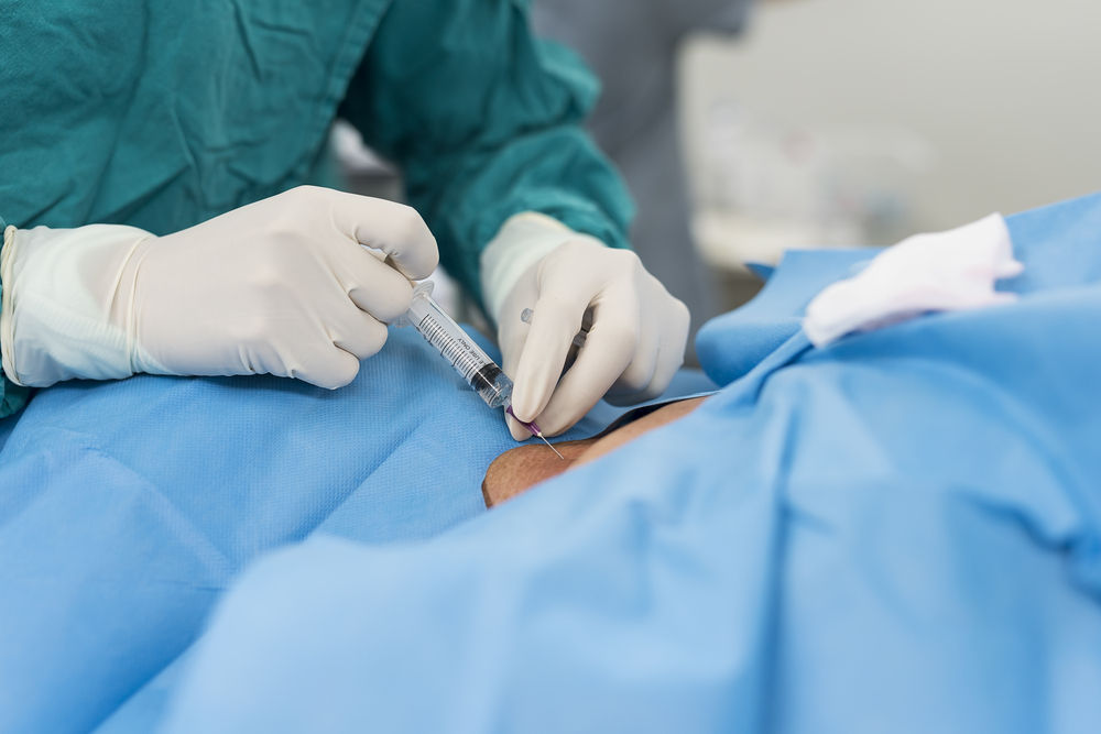
Regional anaesthetic techniques provide excellent anaesthesia and analgesia for many surgical procedures. Regional anaesthesia is gained increased popularity all over the world. Regional anaesthesia is associated with many benefits compared to general anaesthesia, includ- ing reduced morbidity and mortality, superior postoperative analgesia and reduced costs. However, neurological injury after regional an- aesthesia can be distressing to the patients and their families. Most complications of regional anaesthesia are relatively minor, easily man- aged and temporary but in rare cases serious and permanent damage can occur. Neurologic complications associated with central neuraxial block (CNB) can be attributed to incorrect placement of a needle or catheter causing direct neural tissue damage, toxic and infectious agents, and spinal cord compromise resulting from ischemia or mass effect.
Infectious complications
Infectious complications may occur with any regional anaesthetic technique. However those associated with the neuraxial anaesthesia and analgesia may result in devastating morbidity and mortality, in- cluding abscess formation, necrotizing fasciitis, meningitis, arachnoi- ditis, paralysis and death. The risks of serious central or peripheral ner- vous system infection are low, and with strict adherence to sterility during the block performance there is little probability of infection be- ing introduced. However, recent epidemiologic studies suggest the enormous discrepancy of infectious complications associated with neu- raxial techniques. The rate of spinal-epidural abscess or meningitis occurence has been reported to be 1:10 000 to 1:40 000. Aromaa et al. reported lower infectious complication rate of 1.1:100 000 blocks. On the contrary Wang et al. estimated that the incidence of epidural abscess after epidural analgesia was 1:1930 and the risk of permanent neurologic deficit was 1:4343 catheters. This discrepancy can be ex- plained by different data collection techniques, differences in postoper- ative monitoring and reporting and in aseptic technique and antibiotic administration,varying definitions of infection and colonization. The frequency of infection associated with peripheral neural catheters re- mains undefined. Capdevila et al. showed that 29% of peripheral nerve catheters may become colonized with 3% resulting in localized inflammation.
Routes of infection
The etiology of infectious complications is often un- clear. Potential routes might be contaminated syringes, catheter hubs, local anaesthetics or breaches in aseptic technique. Dural puncture has long been considered a risk factor in the pathogenesis of meningitis. The sug- gested mechanisms include introduction of blood into the intrathecal space during needle placement and dis- ruption of the protection provided by the blood-brain barrier.
Skin bacteria gain epidural space through the catheter insertion site. This is supported by Saguraki et al. who traced the source of an epidural abscess to Staphylococ- cus aureus strains fom the patient’s skin flora. Kindleret al. revealed that 83% of 35 epidural catheter related infections, were caused by various Staphylococ- cus species, typical for bacterial flora of the skin.The most frequently detected microorganism on the skin sur- face is S. epidermidis (65–69% of skin flora), while S. aureus (1–2% of skin flora) is the most prevalent micro- organism in epidural infections. This suggests that S. aureus may be more resistant to disinfectants than other microorganisms. However, most epidural abscesses are not related to the placement of indwelling catheters but are believed to be related to infections of the skin, soft tissue, spine or haematogenous spread to the epidural space.
Risk factors
Possible risk factors include underlying sepsis, diabetes, depressed immune status, steroid therapy, localized infection and chronic catheter maintenance. Patients with altered immune status because of neoplasm, immunosuppression after solid organ transplantation and chronic infection with human immunodeficiency virus or herpes simplex virus are often not considered candidates for neuraxial blocks. These patients are susceptible to infection with opportunistic pathogens. Anaesthesiologists have long considered sepsis to be a relative contra- indication to the administration of spinal or epidural anaesthesia. However, if there is a clear benefit to be gained from epidural anaesthesia or analgesia, a septic condi- tion is not an absolute contraindication. Postulated mechanisms for haematogenous infection of the central nervous system caused by subarachnoid or epidural puncture might be an accidental vessel puncture. Antibiotic chemoprophylaxis should be given before the puncture and the patients must be closely followed for the development of epidural abscess. Because of the possibly increased risk of infectious complications, informed con- sent should be obtained from the patient. The decision to perform a regional anaesthetic technique must be made on an individual basis considering the benefits of regional anaesthesia and the risk of central nervous system infection which may occur in any bacteremic patient. Available data suggest that patients with evidence of systemic infection may safely undergo spinal anaesthesia, provided appropriate antibiotic therapy. Meningitis and epidural abscess are both complications of neuraxial blocks, the risk factors and causative organisms are disparate. Staphylococcus is the organism most com- monly associated epidural abscess and often this infections occured in patients with impaired immunity. Meningitis follows dural puncture is typically caused by alphahemolytic streptococci, with the source of the organism the nasopharynx of the anaesthesiologist.
Symptoms, signs and diagnosis
The most frequent signals of an impending epidural infectious complication are local tenderness and infec- tious signs at the skin catheter entry site and new back- ache. Subsequently, increasing leg weakness, loss of sen- sation and sphincter control in the presence of backache are ominous signs. Epidural abscess formation usually occurs days to weeks after neural block associ- ated with fever and leukocytosis. How- ever, it is important to know that an epidural abscess may present with paraplegia and no backache or pyre- xial symptoms.
Computer tomography can help in verifying the diag- nosis, however CT-scans have given false negative find- ings in up to 50% of cases. The most sensitive diag- nostic procedure is MRI with or without gadolinium contrast enhancement.
Treatment of infectious complications
Abscess formation after epidural or spinal anaesthesia can be superficial, requiring limited surgical drainage and intravenous antibiotics, or occur deep in the epidural space with associated cord compression. The latter is for- tunately a rare complication, but it requires aggressive, early surgical decompression to achieve a satisfactory outcome. Antibiotics alone cured some patients who had minimal or no neurological symptoms.