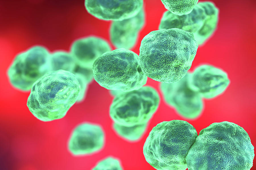
Amebiasis is caused by Entamoeba histolytica , a protozoan that is found worldwide. The highest prevalence of amebiasis is in developing countries where barriers between human feces and food and water supplies are inadequate.
Although most cases of amebiasis are asymptomatic, dysentery and invasive extraintestinal disease can occur. Amebic liver abscess is the most common manifestation of invasive amebiasis, but other organs can also be involved, including pleuropulmonary, cardiac, cerebral, renal, genitourinary, peritoneal, and cutaneous sites. In developed countries, amebiasis primarily affects migrants from and travelers to endemic regions.
E histolytica is transmitted via ingestion of the cystic form (infective stage) of the protozoa. Viable in the environment for weeks to months, cysts can be found in fecally contaminated soil, fertilizer, or water or on the contaminated hands of food handlers.
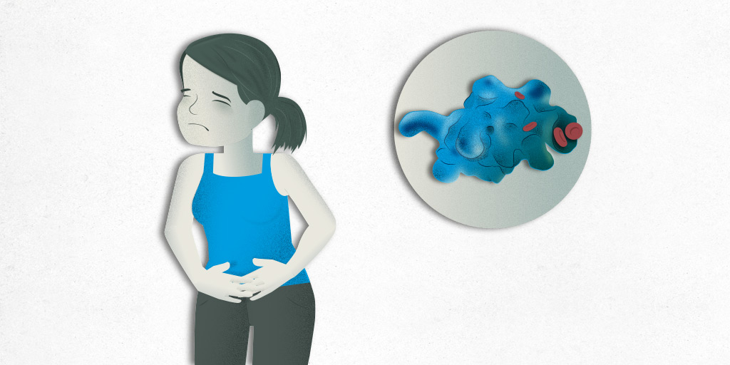
Excystation then occurs in the terminal ileum or colon, resulting in trophozoites (invasive form). The trophozoites can penetrate and invade the colonic mucosal barrier, leading to tissue destruction, secretory bloody diarrhea, and colitis resembling inflammatory bowel disease. In addition, the trophozoites can spread hematogenously via the portal circulation to the liver or even to more distant organs
E histolytica is capable of causing a spectrum of illnesses (see Presentation). Intestinal conditions resulting from E histolytica infection include the following:
Extraintestinal conditions resulting from E histolytica infection include the following:
ETİOLOGY
Amebiasis is a parasitic infection caused by the protozoal organism E histolytica, which can give rise both to intestinal disease (eg, colitis) and to various extraintestinal manifestations, including liver abscess (most common) and pleuropulmonary, cardiac, and cerebral dissemination.
The genus Entamoeba contains many species, some of which (ie, E histolytica, Entamoeba dispar, Entamoeba moshkovskii, Entamoeba polecki, Entamoeba coli, and Entamoeba hartmanni) can reside in the human interstitial lumen. Of these, E histolytica is the only one definitely associated with disease; the others are considered nonpathogenic.
E histolytica is transmitted primarily through the fecal-oral route. Infective cysts can be found in fecally contaminated food and water supplies and contaminated hands of food handlers. Sexual transmission is possible, especially in the setting of oral-anal practices (anilingus). Poor nutrition, through its effect on immunity, has been found to be a risk factor for amebiasis.
PROGNOSİS
Amebic infections can lead to significant morbidity while causing variable mortality. In terms of protozoan-associated mortality, amebiasis is second only to malaria. The severity of amebiasis is increased in the following groups:
Intestinal infections due to amebiasis generally respond well to appropriate therapy, though it should be kept in mind that previous infection and treatment will not protect against future colonization or recurrent invasive amebiasis.
Asymptomatic intestinal amebiasis occurs in 90% of infected individuals. However, only 4%-10% of individuals with asymptomatic amebiasis who were monitored for 1 year eventually developed colitis or extraintestinal disease. [16]
With the introduction of effective medical treatment, mortality has fallen below 1% for patients with uncomplicated amebic liver abscess. However, amebic liver abscess can be complicated by sudden intraperitoneal rupture in 2-7% of patients, and this complication leads to a higher mortality. [4]
Case-fatality rates associated with amebic colitis range from 1.9% to 9.1%. Amebic colitis evolves to fulminant necrotizing colitis or rupture in approximately 0.5% of cases; in such cases, mortality may exceeds 40% [39] or even, according to some reports, 50%.
Pleuropulmonary amebiasis has a 15-20% mortality rate. Amebic pericarditis has a case-fatality rate of 40%. Cerebral amebiasis carries a very high mortality (90%).
DİAGNOSE
A doctor may suspect amebiasis after asking about your recent health and travel history. Your doctor may test you for the presence of E. histolytica. You may have to give stool samples for several days to screen for the presence of cysts. Your doctor may order lab tests to check liver function to help determine if the ameba has damaged your liver.
When the parasites spread outside the intestine, they may no longer show up in stool. So your doctor may order an ultrasound or CT scan to check for lesions on your liver. If lesions appear, your doctor may need to perform a needle aspiration to see if the liver has any abscesses. An abscess in the liver is a serious consequence of amebiasis.
TREATMENT
Treatment for uncomplicated cases of amebiasis generally consists of a 10-day course of metronidazole (Flagyl) that you take as a capsule. Surgery may be necessary if the colon or peritoneal tissues have perforations.
PREVENTİON
Proper sanitation is the key to avoiding amebiasis. As a general rule, thoroughly wash hands with soap and water after using the bathroom and before handling food.