Childhood rashes are cutaneous eruptions of acute onset. Clinically, they may be categorised as maculopapular, pustular, vesiculobullous, diffuse/erythematous, or petechial/purpuric in nature. However, in many aetiologies these forms may co-exist or evolve from one form to another.
Initial considerations in evaluating a rash in children include its morphology, duration, and distribution. Age, sex, family history, medications, known allergies, and exposures are also of primary importance.
Generally, rash in the absence of fever or systemic symptoms is not urgent. However, when fever or signs of illness are present, urgent evaluation and treatment must be considered. The differential diagnosis is extensive, ranging from self-limiting conditions (e.g., roseola) to life-threatening illnesses such as meningococcal disease.
Several systemic conditions with a serious clinical course may have a rash as a component and should be assessed urgently if suspected (see Urgent considerations).
Eczema
This skin rash picture demonstrates a classic case of eczema, which can be helpful in diagnosing your child with eczema.
Eczema is usually diagnosed based on the appearance of the itchy rash in typical areas, including the forehead, cheeks, arms and legs in infants, and the creases or insides of the elbows, knees, and ankles in older children.
Eczema is often described as a very itchy rash, that is often red, rough or irritated, scaly, and can become oozing.
Although eczema can sometimes be hard to control, the basics of preventing eczema can help, including avoiding known triggers, such as harsh soaps, bubble baths, overheating and sweating, wool and polyester clothing, and the liberal use of moisturizers, especially using a moisturizer every day and within 3 minutes of getting out of the bath or shower.
When your child's eczema gets worse or flares, the typical eczema treatments include using topical steroids and the newer non-steroidal medications like Elidel and Protopic.
For hard-to-control eczema, parents might try using an antihistamine to control itching, wet dressings or wet-to-dry dressings, and even antibiotics if your child has signs of a secondary skin infection.
Bug bite
No matter how careful you are about using insect repellents, it is likely that your child will occasionally get a bug bite, such as the one shown below.
The majority of bug bites, whether by insects such as an ant, chigger, or wasp, aren't dangerous, unless your child is allergic to the insect. Even most spider bites, which often resemble regular bug bites, aren't that dangerous unless caused by a black widow or brown recluse spider.
These bug bites can be scary for parents, though, since even a 'normal' reaction to a bug bite, as shown in the picture above, can include redness, swelling, and warm skin.
Some tips to remember about bug bites include that:
Chicken pox
The classic rash of chicken pox infections includes red papules (bumps), vesicles (the spots that look like little blisters), which then become crusted scabs.
Chicken pox typically starts on a child's trunk and then spreads to the rest of their body, including their arms, legs, and head.
Other symptoms of chicken pox typically include a prodrome of fever, malaise, headache, lack of appetite, and mild abdominal pain for 1 to 2 days.
It is very itchy and very contagious but can be prevented with a chickenpox vaccine.
Keep in mind that the current immunization schedule advises that kids get a chicken pox booster shot beginning when they are four years old, which should help to further decrease chickenpox infections.
Fifth disease
The 'slapped cheeks' rash of Fifth Disease is a classic pediatric sign, as can be seen in this picture.
Although many parents dismiss the red cheeks that kids with Fifth Disease get and think it is simple flushing or is caused by sun or wind, when it is followed by the even more classical pattern of getting a pink or red lacelike rash on their arms, the diagnosis is usually easy to make.
Ringworm
The typical ringworm rash on the body looks like a red circular lesion with a scaly border and these areas may be itchy.
An over-the-counter antifungal cream or ointment is the usual treatment for ringworm, except for tinea capitis, which is much more difficult to treat and often requires several months of an oral medication (like Griseofulvin).
Prescription topical creams, suspensions, and lotions are also available, like Loprox, Spectazole and Oxistat are also available.
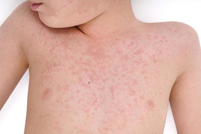
Measles
According to the CDC, 'Measles is an acute, highly communicable viral disease with prodromal fever, conjunctivitis, coryza, cough, and Koplik spots on the buccal mucosa. A characteristic red blotchy rash appears around the third day of illness, beginning on the face and becoming generalized. Measles is frequently complicated by middle ear infection or diarrhea. The disease can be severe, with bronchopneumonia or brain inflammation leading to death in approximately 2 of every 1,000 cases.'
Fortunately, measles can be prevented by the MMR (Measles | Mumps | Rubella) vaccine. Keep in mind that 'measles remains a common disease in many countries of the world, including some developed countries in Europe and Asia.'
Keep in mind that many viral infections cause a 'red blotchy rash,' measles is rare in the United States, especially as most kids are immunized. So unless your child has the pattern of measles symptoms described above, then you likely don't have to worry about measles every time your child gets a rash.
 Anaphylaxis
Anaphylaxis is a life-threatening allergic reaction to foods, insect bites/stings, medications, skin contactants, or inhalants. The cutaneous findings in anaphylaxis include widespread urticaria; swelling of the lips, eyelids, and tongue; and often an erythema or flushing from vasodilation.
The reaction is an immunoglobulin (IgE)-mediated response to an allergen that leads to release of immunological mediators. The reaction can occur immediately after the allergen is introduced or can be delayed. Once the reaction is triggered, it proceeds rapidly.
The most common agents are:
Patients may present with airway compromise, hypotension, and/or tachycardia. Emergency intervention may include giving subcutaneous adrenaline (epinephrine), oxygen, and supportive measures. For children at risk of anaphylaxis, adrenaline auto-injectors should be prescribed in conjunction with a personalised written emergency plan.
TEN and SJS constitute a spectrum of severe generalised exfoliative dermatitis. SJS is milder than TEN, and this is reflected by their respective associated mortality (SJS: 1% to 5%; TEN: 25% to 35%).
Common offending medications include:
Patients may also report or manifest with signs of a recent upper respiratory infection, or infection with mycoplasma, herpes, Epstein-Barr virus, or cytomegalovirus.
There is widespread cutaneous involvement, with associated involvement of ≥2 mucosal surfaces (oral, conjunctival, anogenital). Skin lesions may be initially targetoid (with no central blistering), although they often become confluent. Bullous lesions may also develop, and Nikolsky sign (blister induced with lateral pressure) is noted within affected areas. The lesions are painful and the patient appears acutely ill. Secondary infection may occur.
Initial management includes:
The presentation of DRESS syndrome resembles a morbilliform drug eruption, but the patient is more unwell, often with fever, abdominal pain, and facial swelling. The time interval between intake of the offending medication and symptoms also tends to be longer, often 2 to 6 weeks.
The presence of painful skin lesions, dusky lesions with early erosion, and mucous membrane involvement may signal an evolving erythema multiforme major.
Patients with systemic hypersensitivity syndrome may present with a morbilliform drug eruption. These patients may be acutely unwell, with fever, abdominal pain, facial swelling, and significant lymphadenopathy. Investigations may show elevated transaminases, and a full blood count (FBC) may show marked eosinophilia.
Common offending medications include:
Immediate withdrawal of the medication is indicated. Treatment with oral corticosteroids (e.g., prednisolone) may be required. In severe hypersensitivity reactions that do not respond to prednisolone (prednisone), IVIG can be tried. Mortality in DRESS may be 8% to 10%.
Diagnose
Clinically, a rash may be categorised as maculopapular, pustular, vesiculobullous, diffuse/erythematous, or petechial/purpuric in nature. However, in many aetiologies these forms may co-exist or evolve from one form to another. Diagnosis should focus on exclusion of urgent considerations first, as these are likely to require prompt intervention. Associated symptoms and contact with other children with similar symptoms may help to elucidate the cause.
Initial considerations include morphology, duration, and distribution. Age, sex, family history, medications, known allergies, and exposures are also of primary importance. Patients often reveal the cause of the rash in their historical recollection of events before its appearance (e.g., a mother reports that several children in the neighbourhood who have been unwell with a 'bug' then develop a rash: consider measles).
A full history should include current and previous medications; existing systemic conditions; possible exposure to infection; and social, recreational, and travel information. Contact with people with existing conditions such as impetigo or scabies, and viral infections such as chickenpox, Epstein-Barr virus (EBV), erythema infectiosum (fifth disease), roseola infantum (sixth disease), and hand-foot-and-mouth disease may help to confirm the diagnosis. Any history of pharyngitis (as a source of streptococcal infection) should be noted. Any recent illness associated with a sore throat or upper respiratory tract infection suggests a viral illness. For all viral exanthems, fever, malaise, pharyngitis, and myalgia are commonly associated symptoms.
Examination of the primary lesion should include type of rash, its extent, and distribution. Involvement (or sparing) of the mucous membranes should be noted. A full systemic examination should record any associated features such as pyrexia, pruritus, lymphadenopathy, or hepatosplenomegaly. Generally, rash in the absence of fever or systemic symptoms is not urgent. When fever or signs of illness are present, urgent assessment and treatment must be considered. The differential diagnosis is extensive, ranging from self-limiting conditions (e.g., roseola) to life-threatening illnesses such as meningococcal disease. Several systemic conditions with serious clinical course may have a rash as a component, and these should be assessed urgently if suspected (see below).
For many childhood rashes, diagnosis is clinical, and tests are not routinely recommended. However, laboratory investigations may be appropriate in patients in whom the cause is not clear. In a rash suspected to be caused by a drug, no laboratory tests to determine or confirm the responsible drug are readily available. Full blood count (FBC) may show only mild to moderate peripheral eosinophilia. Metabolic, hepatic, and renal function panels are usually normal. The diagnosis of viral exanthema, depending on the particular virus involved, may need to be confirmed by serologically demonstrable antibody titres, viral culture, or molecular studies (e.g., polymerase chain reaction). Acute hepatitis serology is positive and aminotransferases are elevated in hepatitis B virus (HBV) and hepatitis C virus (HCV) infection. Patients with suspected HIV exanthem will screen positive for HIV viral RNA or core antigen. All patients with more significant clinical disease, including serious bacterial infections, or with extracutaneous findings require a more complete work-up.